
The mechanics of morphogenesis is something European scientists, in particular, seem to find intriguing. However, physical biology is an approach many classical biologists in America have had a difficult time in the past understanding as well as accepting, as evidenced by vociferous attacks in the blogosphere on scientists working in that area. Fortunately, this is changing with America’s new generation of scientists, with project support from organizations like the Simons Foundation, and with publicly funded research in Europe that continues to explore along those lines.
French scientists, in particular, have been central to the inquiry into the mechanics of shape in developmental biology. An inspiring example is the current work of Jean-Léon Maître, who is leading a team at Institut Curie in Paris looking at how the mammalian embryo is built.
Maître is known for his research on the cell “dance” of the early embryo, an investigation he first collaborated on with Takashi Hiiragi a few years ago at the European Molecular Biology Laboratory in Heidelberg, Germany while a postdoc fellow there. Jean-Léon Maître’s PhD studies were at Max Planck Institute for Cell Biology and Genetics and at the Institute of Science and Technology Austria.
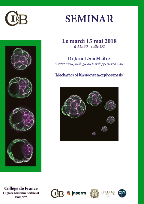
Aside from serving as chef d’équipe of his mammalian embryo lab at Institut Curie, Maître is increasingly in demand as a speaker. In recent months he’s addressed the International Titisee Conference: “From Oocyte to Embryo – Illuminating the Origins of Life” and next week is guest lecturer at Collège de France on the “Mechanics of Blastocyst Morphogenesis.”
I reached Jean-Léon Maître by phone at Institut Curie in Paris several weeks ago for the following conversation.
Suzan Mazur: Would you say mechanics of morphogenesis research is largely coming from Europe?
Jean-Léon Maître: There’s a very strong community in Europe, but there have been many very good projects in the US as well as in Japan. In the US, I can think of Malcolm Steinberg, who proposed very early on [1960s] differential adhesion. It was a landmark study. He was a pioneer in the field of biomechanics. He was at Princeton University. There’s also Ray Keller, a developmental biologist who was working in biomechanics very early on. . . . But it’s true that there’s a fairly large community in Europe, particularly in France actually.
A lot of biomechanics research has been done here at Institut Curie. This came from the efforts of Pierre-Gilles de Gennes, a Nobel laureate in physics who was recognized for his study of liquid crystals. Pierre-Gilles de Gennes started the field of what is now called soft matter, which is not just about biology but about material science in general. His research triggered a great range of studies in biology.
Suzan Mazur: It’s emerging now in a big way, why do you think?
Jean-Léon Maître: It’s emerging in a big way I’m not completely sure. In the field of biology in which I’m working, there’s a huge contribution of mechanics because when the embryo is forming it is not just defining its cell types, it’s defining the biochemistry of the cells depending on where they are and what they need to do. Say, the liver cells have to produce certain types of enzymes and other cells have to produce certain other types of enzymes. But at the same time as you need to define the type of cells, you also need to define the architecture of the embryo. If you want to build something, shape it, carve it, you have to apply forces and consider the mechanical properties of the material you are deforming. So, you need to understand how those forces of mechanics are controlled.
Suzan Mazur: A fair number of classical biologists in the US don’t understand this idea of physical forces and have in the past ridiculed and rejected this approach. Regardless, the science now seems to be moving center stage. It’s interesting.
Jean-Léon Maître: There is a huge contribution that biophysics or physical biology can make to biology. Biology is becoming more and more quantitative and our field is quantitative by nature, so it is really helping to push quantitative measurement into biology in general.
Suzan Mazur: Biology is also becoming increasingly quantitative because of the new tools that are available?
Jean-Léon Maître: Yes, but it’s also a different type of approach. For a very long time biologists were observers. They would observe and describe in a very inquisitive way and that would be it. But it would be sometimes difficult to know whether this observation was a one-time thing or something very common. Quantification has revolutionized chemistry and physics in the past and this is what is happening to biology for a few decades now. It’s really, really, really important what’s happening. Quantification of the field is important.
Suzan Mazur: So better microscopes are available plus computer quantification is revolutionizing biology?
Jean-Léon Maître: Chemistry and physics became quantitative before the advent of computers. Biologists were just too busy with other questions that did not really need quantification. Now it’s becoming [crucial to research]. Without it you might come up with the wrong conclusion.
As for microscopy—yes, there are a lot of new tools for microscopy and biophoto measurement. However, in my lab we work with mammalian embryos, which are very sensitive, very fragile. A fish or a fly in development is exposed to a very rough environment. But mammalian embryos develop inside the mother so they are protected and as a result are much more fragile.
So we use simple microscopes and simple techniques that are actually very old to study these fragile structures. The main tool we use in the lab to measure different mechanical properties is 50 years old. But it works, so that’s fine. . . .
Our microscopes allow us to image our embryos in a much friendlier way. By using much less light we collect more information.
Suzan Mazur: Why does the field have so many names—new condensed matter physics, soft matter, mechanics in morphogenesis, morphomechanics, geometric morphometrics, etc.?
Jean-Léon Maître: That’s something we are often discussing. I am a biologist, I was trained as a biologist and I learned a little bit of physics during my PhD. But I am a biologist. We have trouble sometimes defining the field name.
It’s obviously biophysics because we’re using physical tools to study biology, but the problem is that biophysics also encompasses structural biology. So scientists who specialize in x-ray technology—MRI, etc., they are also biophysicists. But we don’t do the same thing at all. So we are trying to distinguish the different areas of research by using different names. Physical biology is one that I like. But there’s no consensus on that.
Suzan Mazur: How many labs would you say are working along similar lines as yours worldwide?
Jean-Léon Maître: Counting only the labs working in developmental biomechanics, maybe 100. . . . It’s a pretty rough estimate. I really don’t know.
Suzan Mazur: Would you briefly describe your work?
Jean-Léon Maître: Our research focus is on how the early mammalian embryo is developing. The early mammalian embryo forms a structure called the blastocyst. . . .
The blastocyst has a specific architecture. The part we’re really trying to understand is how this architecture is shaped and how the embryo is built. What are the forces—pulling the cell this way or that way so that it has the correct shape.
Suzan Mazur: Would you describe the “dance” you’ve observed in embryonic development—the cyclic contractile events—and say how you think this has been conserved in evolution.
Jean-Léon Maître: To understand this we look at cell behavior that is able to deform cells and tissues. One of the major, major, major engines of animal development is contractility of the actin and myosin cytoskeleton. Just like in your muscles you have actin and myosin and fibers that can contract your muscles—many animal cells can also contract in this way. This engine is able to deliver forces that can bend and deform the embryo. This is a major morphogenetic engine conserved in evolution.
If you look at the tissues of developing animals, you will find very, very, very often that what is shaping the tissues is the actin-myosin cytoskeleton. It turns out that this engine that is conserved in evolution in animals, it has a peculiar way of acting. Instead of contracting in a steady way like you could contract your muscles if your try to lift a cup with your hand, the cells do not seem to deform the embryo in a steady way but rather in a pulsatile way.
This is true in worms, this is true in Drosophila, in fish, in starfish, in zebrafish. What we saw is that we have the very same kind of pulsatile contraction in the early mouse embryo. And in the early mouse embryo those pulses of contraction take a very particular form, which is one of a wave of contraction that physically travels around the cell perimeter in a periodic fashion. It goes around the cell every 80 seconds. [J-L Maître calls this “pecowaco,” short for periodic cortical wave of contraction.]
What is also very germane is that the period of oscillation in those mammalian cells is very similar to what you can find in fish, starfish, zebrafish, and to some extent in Drosophila and in worms. About every minute to 100 seconds there is pulsation [a “dance” because a wave of contraction is bending the cell’s surface].
Suzan Mazur: And why do you think these cyclic contractile events have been conserved in evolution?
Jean-Léon Maître: This short answer is, I have no idea. What the field is leaning towards is that this constraint on the time scale of contraction is some kind of structural constraint. The way the actin cytoskeleton and myosin forces are organized at the molecular level kind of defines the time scale.
Suzan Mazur: Do viruses have anything to do with this conservation?
Jean-Léon Maître: No, I don’t think so, I don’t see any hint of why viruses would be involved. What I find really interesting, though, is that the time scale of the development—let’s say of a worm or of a fly or a fish embryo or of a mouse embryo—are very, very different. And humans are developing on a time scale of days, weeks, months. Whereas animals used in the lab, like flies, develop super-fast, so the time scale of the deformation of those embryos is very close to the time scale of those periodic contractions.
With slower developing embryos, we have the same kind of rhythm in terms of motion and tension but the embryo itself deforms in a very different time scale. How those two time scales are talking to each other is something I think is very, very poorly understood. . . .
Suzan Mazur: Thank you. How common across species is the blastocyst?
Jean-Léon Maître: The blastocyst is a structure of the early mammalian embryo before it implants. If you look across mammals—like in mouse, human, cow, or if you go all the way to marsupial—each will form a cyst, a spherical fluid-filled structure with an inner cell mass and outer layer of cells. . . . All mammals form a blastocyst, an embryo. There are other embryos, such as sea urchin embryos, that also form a cyst in their very early development. So it is not a structure specific to mammals only.
Suzan Mazur: But the blastocyst is not common to animals outside of mammals.
Jean-Léon Maître: Right, although many animals in the embryonic stage form fluid-filled organs, blood vessels that are fluid-filled organs. This is very common.
Suzan Mazur: Is surface tension the most pronounced physical force driving compaction in embryonic development?
Jean-Léon Maître: Yes.
Suzan Mazur: Are you part of the 2018-2023 ERC Mechablasto project, and if so, what is your role in the project?
Jean-Léon Maître: Yes. I have my own research group. I proposed a project to the funding agencies and the project got funded. I am running this project.
Suzan Mazur: You study mammalian embryonic development. But plants have embryos as well. A similar inversion happens with algae, for instance. Are some of the same mechanisms apparent in plant embryonic development as in mammalian development?
Jean-Léon Maître: It depends on what you mean by mechanisms. The mechanisms are very different in plants and animals. . . . As I mentioned, in animals the actin-myosin cytoskeleton is important. In plants that’s not the case. In plants hydrostatic pressure builds in the cytoplasm and it pushes, in a directed way, by changing the resistance of the walls in an oriented fashion. Different mechanisms in plants and animals, but the forces are the same. If you want to bend something, you just have to push on it or pull on it.
Suzan Mazur: Thank you. Jack Szostak has said understanding self-organization is crucial to protocell development and it is not yet really understood. Do you see protocell development as technically feasible?
Jean-Léon Maître: My background has very little to do with that. At some point the first cell appeared through some self-organized process. So, yes, I guess so.
Suzan Mazur: What role do you recognize for viruses in embryonic development? You said earlier that you didn’t think that viruses were involved in conserving the cyclic contractile events.
Jean-Léon Maître: This is not really my expertise either but I know from the scientific literature that viruses have left in the human genome some genes that are important in implantation of the human embryo.
Suzan Mazur: You were a postdoctoral researcher in Takashi Hiiragi’s group at European Molecular Biology Laboratory in Heidelberg. Do you have anything to say about that experience?
Jean-Léon Maître: Takashi Hiiragi is someone who is very open, he is curious to explore new directions. It was really pleasant to work with him. Ours was a very natural collaboration. We were working together on the mouse embryo.
Suzan Mazur: I think it was Hiiragi who commented at the Princeton “Mechanics in Morphogenesis” event in February—which was streamed over the Internet—about the need to see cells as 3D balloons, not 2D. Do you have any reservations about reliance on computer modeling and computer simulation that’s going on in your field?
Jean-Léon Maître: Unfortunately, we are limited to 2D for much of the modeling we are doing because the equations we use for 3D are not solvable. So to a large extent we are stuck with 2D if we want to make an analytical model, which is a nice pretty equation where we can understand the terms and what happens when we change one of the terms. If you want to go 3D—which is very important, of course—those 3D models will be simulations. We are not exploring all the possibilities, only a subset of the space of those equations. Because those equations are just too complicated to find one answer.
So with 2D we can make a very elegant map that can be used to model development. With 3D we cannot be as thorough. So we use them both. When we can, we use 3D; when we cannot, we use 2D. I think those two approaches are very complementary.
Suzan Mazur: Is there a final point you’d like to make?
Jean-Léon Maître: Yes. One area we are not exploring enough in our field is the originary aspect of the mechanics of shape. How changes in forces and force patterns in different embryos can give rise to diversity, which can eventually give rise to different species. That’s something that would be very interesting to look into.
